Tibial Tubercle Transfer and Medial Patella- Femoral Ligament Reconstruction
Patellofemoral Instability – Understanding the Condition and Treatment Options
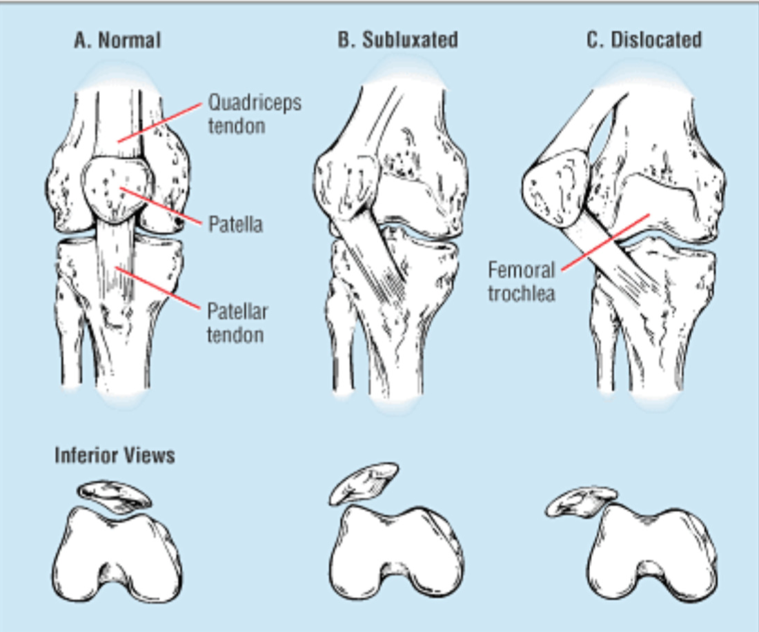
Patellofemoral instability is a common cause of knee pain and disability. It occurs when the kneecap (patella) does not track properly within the groove of the thigh bone (trochlea), leading to episodes of subluxation or dislocation. Most dislocations occur laterally (towards the outside).
Symptoms
- A feeling that the knee “gives way”
- Recurrent dislocations or subluxations
- Pain at the front of the knee, especially during activities like climbing stairs, running, jumping, or changing direction
Risk Factors
- Shallow trochlear groove (trochlear dysplasia)
- Patella alta (high-riding kneecap)
- Increased Q-angle or knee valgus
- Weak medial stabilizers (VMO muscle)
- Previous injury to the medial patellofemoral ligament (MPFL)
- Abnormal position of the tibial tubercle
Initial Management
Treatment usually begins with non-operative measures:
- Physiotherapy to strengthen stabilizing muscles
- Bracing or taping
- Activity modification
However, if instability persists despite adequate rehabilitation, surgery may be necessary to prevent recurrent dislocations and reduce the risk of developing arthritis in the patellofemoral joint.
Surgical Options – A Tailored Approach
Surgery for patellofemoral instability is not one-size-fits-all. It often involves a combination of procedures, depending on the underlying cause:
- MPFL Reconstruction (MPFLR) – Restores the primary soft tissue restraint.
- Tibial Tubercle Transfer (TTT) – Realigns the patella by repositioning the tibial tubercle (where the patellar tendon attaches).
What Does TTT Involve?
- The tibial tubercle is carefully cut from the front of the tibia, shifted to improve alignment, and fixed in its new position.
- Because the bone is cut, it requires time to heal:
- Immobilization in a splint for 6 weeks
- Full recovery can take up to 3 months
Why Choose A/Prof Hazratwala?
A/Prof Hazratwala performs these procedures regularly with predictable, reliable outcomes. His approach is based on restoring normal anatomy and biomechanics to achieve long-term stability and prevent future arthritis.
Bottom line: If you suffer from recurrent patellar dislocations or persistent instability, early surgical intervention can protect your knee from long-term damage. Book a consultation to discuss whether MPFL reconstruction, tibial tubercle transfer, or a combination of procedures is right for you.

Sub-Menu
- Queensland Lower Limb Clinic Procedures
- Adult Total Hip Replacements
- Hip Resurfacing Arthroplasty
- Adult Total Knee Replacements
- Adult Revision Hip And Knee Replacements
- Unicompartmental Knee Replacement
- Anterior Cruciate Ligament Reconstruction
- Ankle Reconstruction
- Foot Disorders
- HTO (High Tibial Osteotomy)
- Lower Limb Trauma
- OATS (Osteochondral Autologous Transplantation Surgery)
- Trochanteric Bursitis Surgery
- Knee Arthroscopy
- Surgery for Patella Instability
- Bone Tendon Bone Allograft ACL Reconstruction